The brain computes under constraint. Every dendritic integration event, every synaptic change, and every organelle repositioning reflects a solution to the problem of adaptive function within limits on energy, space, and molecular machinery.
My research asks: what are those limits, how do they shape computation and learning, and when do they become the conditions for failure? I work at the intermediate scale, sitting between molecular mechanisms and systems-level behavior, developing computational models that are biophysically grounded, analytically interpretable, and testable against existing datasets.
Why the intermediate scale?
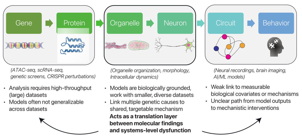
Why this scale? I study cellular organization because morphology, organelle dynamics, and synaptic structure provide measurable links between molecular mechanisms and systems-level function or dysfunction.
At this scale, structural organization becomes a predictive scaffold: it can connect molecular and cellular mechanisms to a neuron’s computational role, its capacity to adapt, and its vulnerability under stress.
How the pieces fit
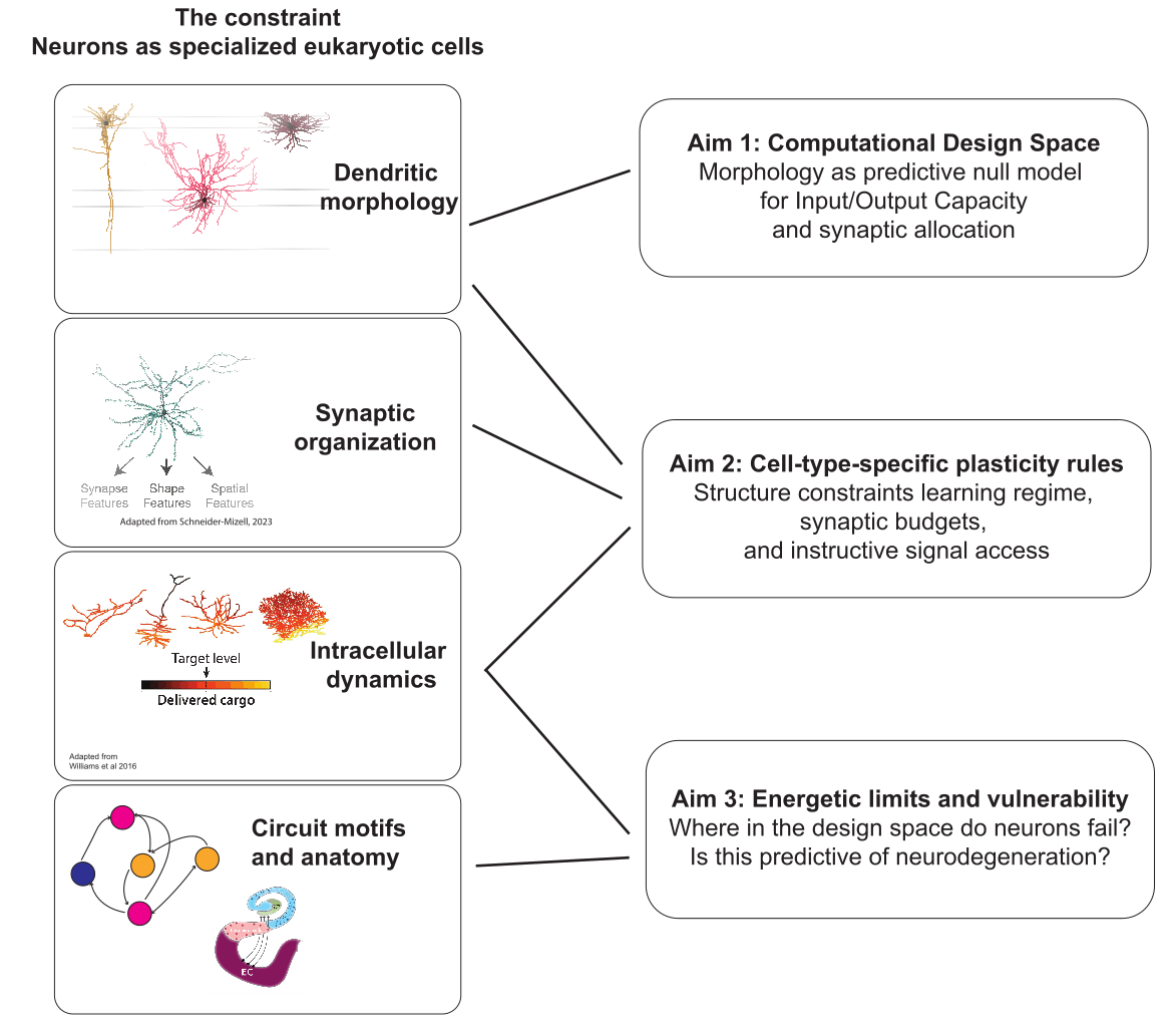
Select an aim in the figure to see how it connects to the work below.
Neuronal Morphology and Computational Complexity
How much does the shape of a neuron determine what it can compute?
Neurons are not simple threshold units. Their dendrites branch, compartmentalize, and integrate inputs in spatially structured ways, and that structure is not incidental. In this line of work, I’ve developed scalable models that treat dendritic architecture as a formal constraint on computational complexity, asking which classes of input-output functions a neuron can learn given its morphology, and how efficiently it can adapt between tasks.
The key finding: branching structure places quantifiable bounds on a neuron’s computational capacity, and the geometry of those branches - whether shallow and broad, or deep and hierarchical - determines not just what a neuron can compute, but how robustly it can do so. Apical-like and basal-like architectures turn out to learn qualitatively different sets of functions, with distinct tradeoffs in generality and retraining speed.
Agrawal A, Buice MA (2025). Bounds on the computational complexity of neurons due to dendritic morphology. NeurIPS 2025. Conference paper
Bayesian Inference of Neurodegenerative Progression
How does Alzheimer’s disease spread, which cells are most vulnerable, and can we infer this from cross-sectional human data?
In collaboration with Mariano Gabitto and Gonzalo Mena (Allen Institute / CMU), I developed B-BIND (Biophysical Bayesian Inference for Neurodegenerative Dynamics): a generative model that reconstructs continuous disease trajectories from snapshots of human neuropathology data, embedding physical priors on protein aggregation, transport, and cell density. The framework infers a pseudotemporal disease axis without requiring longitudinal data that is ethically and practically impossible to collect from human brain tissue.
B-BIND was first applied to the Seattle Alzheimer’s Disease Brain Cell Atlas (SEA-AD), an integrated multimodal atlas of Alzheimer’s disease in humans, to model vulnerability patterns of neuronal and non-neuronal cells in the middle temporal gyrus. The framework is now being extended across the brain: a recent SEA-AD consortium preprint applied an updated version of B-BIND to the caudate nucleus, incorporating two independent processes to capture the distinct dynamics of amyloid and tau pathology in subcortical regions. Multiregion extensions are ongoing.
Agrawal A*, Rachleff VM, Travaglini KJ, et al. (2026). B-BIND: Biophysical Bayesian inference for neurodegenerative dynamics. The Annals of Applied Statistics, 20(1), 285-306. Journal article
Gabitto MI, Travaglini KJ, Rachleff VM, …, Agrawal A, …, et al. (2024). Integrated multimodal cell atlas of Alzheimer’s disease. Nature Neuroscience. Article
Kana OZ, Postupna N, Agrawal A, et al. (2026). The Caudate Nucleus Exhibits Distinct Pathology and Cell Type-Specific Responses Across Alzheimer’s Disease. bioRxiv. Preprint
Intracellular Transport and Metabolic Resilience
(PhD work, UC San Diego, Elena Koslover lab)
How do neurons - cells that can extend millimeters or even meters - maintain metabolic stability in their most distal branches?
My PhD research used analytical and agent-based models to study how mitochondrial transport and turnover act as a distributed control system for neuronal energy supply. The key insight is that mitochondrial dynamics (transport/fusion/fission/autophagy) and their associated regulatory mechanisms couple local metabolic demand and supply to organelle positioning in ways that allow neurons to sustain activity even at large distances from the soma.
In collaboration with Erin Barnhart’s lab (Columbia), we extended these models to Drosophila neurons and showed that dendritic geometry alone can enforce equitable mitochondrial distribution across branches, with structure doing the work that active regulation might otherwise require.
Donovan EJ*, Agrawal A*, et al. (2024). Dendrite architecture determines mitochondrial distribution patterns in vivo. Cell Reports, 43. Article
Agrawal A, Koslover EF (2021). Optimizing mitochondrial maintenance in extended neuronal projections. PLoS Computational Biology, 17, e1009073. Article
Agrawal A, Pekkurnaz G, Koslover EF (2018). Spatial control of neuronal metabolism through glucose-mediated mitochondrial transport regulation. eLife, 7, e40986. Article